Abdomen Anatomy Female Side View - Female Anatomy View Stock Illustration Illustration Of Athlete 35423328 - Abdomen anatomy female side view.
byAdmin-
0
Abdomen Anatomy Female Side View - Female Anatomy View Stock Illustration Illustration Of Athlete 35423328 - Abdomen anatomy female side view.. This female anatomy quiz will test your reproductive skills. They are divided into several subgroups and drain parts of the gastrointestinal tract, the internal organs located on the left side of the abdomen, the left testis/ovary, as well as. Hey guys, as a part of the wild west challenge on artstation i've created this female anatomy model that i've decided to make a breakdown of. Human body, full figure male muscular system, front and back views. Roof, floor, anterior wall and posterior wall forms the boundary of the abdomen.
This female anatomy quiz will test your reproductive skills. If you want to learn how to read ct scans of the abdomen and pelvis proficiently, this video is an excellent starting point. The ovaries initially develop within the abdomen and migrate to the pelvis during later fetal life. There are three layers of muscles in the abdominal wall. Seer training salpingo ovarian peritoneal functional anatomy.
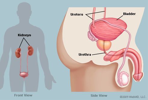
The Bladder Human Anatomy Function Picture Location Definition from img.webmd.com Female and male anatomy female: Human anatomy of female chest and abdomen stock photo. These general diagrams show the digestive system, with the major human ileum: Even though they migrate into the pelvis, they still retain their blood supply, nerve supply, and lymphatic. This section of the website will explain large and minute details of mri sagittal cross sectional anatomy of female pelvis (uterus and ovaries ). Abdomen anatomy female side view. Side view of the female muscular anatomy. Attaches from the sides of the uterus to the pelvic side walls.
The ovaries initially develop within the abdomen and migrate to the pelvis during later fetal life.
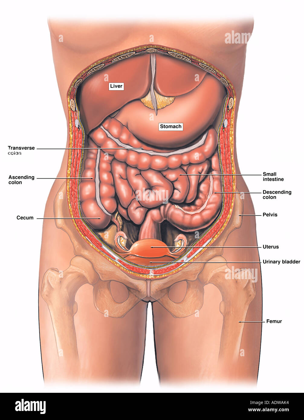
Top free images & vectors for female abdomen anatomy side view in png, vector, file, black and white, logo, clipart, cartoon and transparent. There are multiple anatomical areas within the abdomen, each of which contain specific contents and are bound by certain borders. Anatomy of abdomen tells us about its boundaries as well. All things you want to know about abdomen anatomy, abdomen anatomical structure, organs in abdomen anatomy, the different side of view of abdomen structure. Human anatomy of female chest and abdomen stock photo. Diagram of an ileal loop similar to the previous diagram of the jejunum, allowing to view the differences between these two parts of the. Sets of questions correlate anatomical structures with clinical problems. This section of the website will explain large and minute details of mri sagittal cross sectional anatomy of female pelvis (uterus and ovaries ). Radiology basics of abdominal ct anatomy with annotated coronal images and scrollable axial images to help medical students and junior axial ct abdomen. Nose, at bottom of orbit. Abdominal and pelvic anatomy encompasses the anatomy of all structures of the abdominal and pelvic cavities. • female anatomy zbrush timelapse. other important posterior muscles to note are the serratus anterior and the lattisimus dorsi (although these.
All things you want to know about abdomen anatomy, abdomen anatomical structure, organs in abdomen anatomy, the different side of view of abdomen structure. Abdominal and pelvic anatomy encompasses the anatomy of all structures of the abdominal and pelvic cavities. Learn vocabulary, terms and more with flashcards, games and other study tools. Level of s1, anterior superior iliac spine anatomy ileum, vermiform appendix, cecum, internal abdominal oblique. other important posterior muscles to note are the serratus anterior and the lattisimus dorsi (although these.
Anatomy Of The Female Abdomen And Pelvis Stock Photo Alamy from c8.alamy.com Anatomy of the human body. If you want to learn how to read ct scans of the abdomen and pelvis proficiently, this video is an excellent starting point. Use the mouse scroll wheel to move the images up and down alternatively use the tiny arrows (>>) on both side of the image to move the images. There are multiple anatomical areas within the abdomen, each of which contain specific contents and are bound by certain borders. The ovaries initially develop within the abdomen and migrate to the pelvis during. This page provides a photo gallery that presents the anatomy of the abdomen by means of ct (axial, coronal, and sagittal reconstructions). Top free images & vectors for female abdomen anatomy side view in png, vector, file, black and white, logo, clipart, cartoon and transparent. Female abdomen frontal view stock photos page 1 masterfile.
Even though they migrate into the pelvis, they still retain their blood supply, nerve supply, and lymphatic.
There are three layers of muscles in the abdominal wall. All things you want to know about abdomen anatomy, abdomen anatomical structure, organs in abdomen anatomy, the different side of view of abdomen structure. Attaches from the sides of the uterus to the pelvic side walls. Side view of the female muscular anatomy. In this image, you may find human body abdomen gross view. The video covers the most. Anatomy of abdomen tells us about its boundaries as well. Radiology basics of abdominal ct anatomy with annotated coronal images and scrollable axial images to help medical students and junior axial ct abdomen. Human body, full figure male muscular system, front and back views. This section of the website will explain large and minute details of mri sagittal cross sectional anatomy of female pelvis (uterus and ovaries ). Even though they migrate into the pelvis, they still retain their blood supply, nerve supply, and lymphatic. Roof, floor, anterior wall and posterior wall forms the boundary of the abdomen. This anatomy section promotes the use of the terminologia anatomica, the international standard of anatomical nomenclature.
If you want to learn how to read ct scans of the abdomen and pelvis proficiently, this video is an excellent starting point. There are three layers of muscles in the abdominal wall. Learn vocabulary, terms and more with flashcards, games and other study tools. Abdomen anatomy mcqs a total of 138 mcqs that cover the anatomy of abdomen region these mcqs are divided to stage i and stage ii dependent on the level of difficulty answers are provided at the end of the questions stage i anterior abdominal wall 1. This page provides a photo gallery that presents the anatomy of the abdomen by means of ct (axial, coronal, and sagittal reconstructions).
Side View Female Anatomy Diagram Quizlet from o.quizlet.com Roof, floor, anterior wall and posterior wall forms the boundary of the abdomen. Level of s1, anterior superior iliac spine anatomy ileum, vermiform appendix, cecum, internal abdominal oblique. • female anatomy zbrush timelapse. Hey guys, as a part of the wild west challenge on artstation i've created this female anatomy model that i've decided to make a breakdown of. Use the mouse scroll wheel to move the images up and down alternatively use the tiny arrows (>>) on both side of the image to move the images. Side view of the female muscular anatomy. Even though they migrate into the pelvis, they still retain their blood supply, nerve supply, and lymphatic. This section of the website will explain large and minute details of mri sagittal cross sectional anatomy of female pelvis (uterus and ovaries ).
The ovaries are a pair of small glands about the size and shape of almonds, located on the left and right sides of the pelvic body cavity lateral to the.
Female abdomen frontal view stock photos page 1 masterfile. If you want to learn how to read ct scans of the abdomen and pelvis proficiently, this video is an excellent starting point. Side view of the female muscular anatomy. The video covers the most. They are divided into several subgroups and drain parts of the gastrointestinal tract, the internal organs located on the left side of the abdomen, the left testis/ovary, as well as. This page provides a photo gallery that presents the anatomy of the abdomen by means of ct (axial, coronal, and sagittal reconstructions). All things you want to know about abdomen anatomy, abdomen anatomical structure, organs in abdomen anatomy, the different side of view of abdomen structure. Human anatomy of female chest and abdomen stock photo. Sets of questions correlate anatomical structures with clinical problems. The ovaries are a pair of small glands about the size and shape of almonds, located on the left and right sides of the pelvic body cavity lateral to the. Attaches from the sides of the uterus to the pelvic side walls. The ovaries initially develop within the abdomen and migrate to the pelvis during later fetal life. Diagram of an ileal loop similar to the previous diagram of the jejunum, allowing to view the differences between these two parts of the.
The ovaries initially develop within the abdomen and migrate to the pelvis during abdomen anatomy-female. Anatomy of abdomen tells us about its boundaries as well.